Information
The piriformis muscle originates from the front of the sacrum, the triangular bone situated between the hip bones in the pelvic region. Extending across the sciatic nerve to the top of the femur, the piriformis plays a crucial role in facilitating side-to-side movement of the thigh. Should a spasm occur in the piriformis muscle, it can exert pressure on the sciatic nerve, leading to the manifestation of symptoms collectively known as piriformis syndrome.
The sciatic nerve, a lengthy and robust nerve in the body, either runs alongside or traverses through the piriformis muscle. From there, it descends down the back of the leg, eventually branching off into smaller nerves that terminate in the feet. Piriformis muscle spasms have the potential to compress the sciatic nerve, causing nerve-related symptoms.
Piriformis block induces symptoms akin to genuine sciatica, resulting from the irritation of the sciatic nerve by the piriformis muscle. Commonly referred to by alternative names such as wallet neuritis and deep gluteal syndrome, it manifests as a distinct set of symptoms associated with the compression or irritation of the sciatic nerve by the piriformis muscle.
The primary focus of treating piriformis syndrome is to alleviate buttock and leg pain, stabilize the hip and thigh, and enhance mobility in the hip. Initially, nonsurgical approaches are often explored, with rest, physical therapy, and medication being common first-line treatments. In cases where oral medication fails to provide sufficient pain relief, injection treatments may be recommended. Surgical intervention for piriformis syndrome is rarely considered, and it becomes a viable option only when the underlying cause is severe or progressive neurological deficits, such as leg weakness, are present.
Nonsurgical treatments for piriformis syndrome typically encompass a combination of strategies, including activity modifications (such as avoiding prolonged sitting), posture training, and engaging in physical therapy. In mild cases, taking a rest for 1-2 days may be beneficial in alleviating symptoms.
Symptoms
The symptoms and signs of piriformis syndrome can vary from mild discomfort to severe, debilitating pain in the buttock. If the piriformis muscle exerts pressure on the sciatic nerve, these symptoms may extend down the thigh and leg.
Piriformis syndrome symptoms may exhibit a consistent presence throughout the day or intensify during physical activities involving movements of the hip and legs. Common triggers include sitting, walking, climbing stairs, and twisting. The range of symptoms underscores the dynamic nature of the condition, which can manifest as a spectrum from mild discomfort to more severe and restrictive pain based on the level of piriformis muscle involvement and its impact on the sciatic nerve.
Common Symptoms of Piriformis Block
The common symptoms and signs associated with piriformis syndrome may include a combination of the following:
Buttock pain: Piriformis syndrome typically causes deep buttock pain, described as aching, burning, throbbing, or shooting. This pain often exacerbates after physical activities such as sitting, walking, running, or cycling, as well as activities involving repetitive hip and thigh motion. Prolonged periods of inactivity, such as sitting at a desk or driving, can also worsen the pain.
Numbness and tingling: Numbness and tingling sensations may be experienced in the buttock and down the leg on the affected side. These sensations can intensify during activities like sitting or standing for extended periods.
Difficulty sitting: Pain and tenderness in the buttock may make it challenging to sit for extended periods, especially on hard or uneven surfaces.
Weakness or difficulty moving the leg: The leg on the affected side may feel heavy or stiff, and there may be difficulty lifting it or moving it in specific directions that put tension on the piriformis muscle.
Less Common Symptoms of Piriformis Block
Less commonly, individuals with piriformis syndrome may experience the following symptoms and signs:
Bilateral piriformis syndrome: In fewer than 10% of cases, symptoms may occur simultaneously in both the right and left buttocks and legs.
Alternating symptoms: Rarely, piriformis muscle pain may alternate between both legs. This alternating pattern may suggest the coexistence of degenerative issues in the sacroiliac (SI) joint along with piriformis syndrome.
Symptoms after pregnancy: Piriformis syndrome may develop months after pregnancy, attributed to the elongation and strain of gluteal muscles in the pelvis.
Groin pain and numbness: Piriformis syndrome can lead to groin pain and numbness due to the impingement of the pudendal nerve. This nerve passes below the piriformis muscle and supplies the groin area. It's essential to note that groin pain and numbness may also be associated with a severe but rare medical condition called cauda equina syndrome—a medical emergency requiring immediate treatment.
Any concerning symptoms in the lower back, buttock, pelvis, or thigh should be discussed with a doctor for an accurate diagnosis and prompt treatment. Additionally, symptoms such as groin pain and numbness warrant careful evaluation to rule out serious conditions like cauda equina syndrome.
Diagnosis
Diagnosing piriformis block can indeed be challenging due to the similarity of its symptoms with other conditions like herniated discs or sacroiliac joint dysfunction. To achieve an accurate diagnosis, healthcare professionals, especially those with specialized training in musculoskeletal disorders such as physiatrists, sports medicine specialists, and orthopedic specialists, play a crucial role. The diagnostic process involves several key steps:
Physical examination: A thorough physical examination is conducted in the doctor's office, encompassing clinical tests that assess the range of motion, muscle strength, and reflexes in the lower back and legs. Physicians look for signs of muscle tenderness or inflammation and perform specific tests to evaluate the strength and function of the piriformis muscle and the sciatic nerve.
Review of medical history: Understanding the individual's medical history is essential. This includes detailed inquiries about the onset and characteristics of symptoms, factors that exacerbate or alleviate symptoms, and any other relevant information that may aid in the diagnosis.
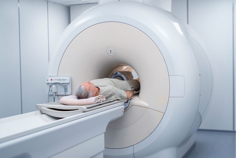
Imaging studies: To assess the health of bones, joints, and soft tissues in the lower back and pelvis, imaging studies such as x-rays, magnetic resonance imaging (MRI), or computerized tomography (CT) scans may be ordered. These studies help identify structural abnormalities or degenerative changes contributing to the symptoms.
In some cases, a referral to a specialist may be necessary for further evaluation and treatment. Specialists such as physiatrists, physical therapists, neurologists, or orthopedic surgeons can provide additional expertise and guidance in managing piriformis syndrome. Collaborative efforts between healthcare professionals are often instrumental in ensuring a comprehensive and accurate diagnosis, leading to effective treatment strategies.
Imaging Tests and Nerve Stimulation
Diagnostic testing beyond a medical history and physical exam serves primarily to rule out other potential causes of buttock pain or leg pain. These tests aim to identify structural abnormalities or degenerative changes contributing to the symptoms. Some of the commonly employed tests include:
Electromyography (EMG): EMG is a minimally invasive test that measures the electrical activity produced in a muscle in response to nerve stimulation. It helps identify muscle dysfunction in the buttock, including the piriformis and surrounding muscles. In piriformis syndrome, abnormal patterns may be observed in the gluteus maximus and piriformis muscles.
Imaging tests: Conditions with symptoms resembling piriformis syndrome, such as bone spurs or herniated discs, can be detected through imaging tests like MRI or CT scans. An MRI scan may reveal anatomical changes within the piriformis muscle, aiding in the diagnosis. Magnetic Resonance Neurography (MRN) may be used to assess the health of nearby nerves.
Ultrasound: Diagnostic ultrasound can analyze the thickness and cross-sectional area of the piriformis muscle, contributing to the diagnosis of piriformis syndrome. This test may be used alone or in conjunction with CT or MRI, and it does not involve radiation exposure, making it preferable in certain situations, such as for patients with implants.
The combination of clinical tests, nerve stimulation, and imaging studies conducted by a physician helps rule out other conditions that may mimic piriformis syndrome, facilitating a more accurate diagnosis and informing the most suitable treatment approach.
Once piriformis syndrome is diagnosed, a structured treatment plan is formulated. Both nonsurgical and surgical treatments may be employed based on the severity of the condition and the individual's response to initial interventions.

Procedure
In the initial stages of treating piriformis syndrome, nonsteroidal anti-inflammatory drugs (NSAIDs) and muscle relaxants are commonly prescribed. These medications aim to alleviate pain and reduce muscle tension.
Physical therapy plays a pivotal role in managing piriformis syndrome, as focused exercises can contribute significantly to resolution. Therapeutic modalities such as Ultrasound massage and Deep Tissue Release techniques are employed, accompanied by a structured program that incorporates specific stretches for the piriformis and hamstring muscles.
For patients who do not respond adequately to conservative measures, additional options are available. Ultrasound-guided (USG) or Fluoroscopy-guided injections, comprising local anesthetics (LA) and steroids, can be administered to target the affected area and provide relief.
BOTOX injections also present a viable option, as they induce functional denervation of the muscle, effectively relieving spasm and associated symptoms.
In cases where conservative and injectable treatments prove insufficient, surgical interventions may be considered. Surgical options include the release of the tendon at its attachment on the greater trochanter, dissection through the piriformis muscle, or neurolysis of the sciatic nerve. These procedures aim to address the underlying issues contributing to piriformis syndrome, providing a more definitive solution for those who have not found relief through non-surgical interventions.
Surgical interventions for piriformis syndrome encompass the following procedures:
Piriformis muscle release: This surgery is designed to alleviate compression on the sciatic nerve by cutting the tendon that attaches the piriformis muscle to the top of the thigh bone (femur).
Sciatic nerve decompression: This surgical approach aims to relieve sciatic nerve compression and alleviate pain caused by the inflamed piriformis muscle. It involves removing a small portion of the muscle that compresses the nerve, thus reducing pressure on the nerve.
Endoscopy: Endoscopic surgery, a minimally invasive method, may be considered for piriformis syndrome patients. It has the potential to yield better outcomes with fewer complications compared to open surgical approaches.
As with many surgical procedures, potential risks such as bleeding, nerve damage, and infection should be acknowledged in piriformis surgeries. Endoscopic surgery for piriformis muscle release is deemed minimally invasive and elective, allowing for pre-planned scheduling with the surgeon rather than necessitating urgent or emergent intervention.
It's crucial to recognize that surgery does not guarantee a complete resolution of symptoms. Patients contemplating surgery should engage in thorough discussions with their surgeon to understand potential benefits, risks, and explore alternative surgical options before making an informed decision.