Information
What is Discography?
A discography, alternatively known as discogram, serves as an imaging test designed for the assessment of back pain. This diagnostic procedure aids your physician in determining whether a specific abnormal disc within your spine is the root cause of your back pain.
Spinal discs function as spongy cushions situated between the vertebrae (bones) of the spine. In the course of a discogram, a contrast dye is injected into the soft center of one or more discs. Notably, this injection may replicate the sensations of your back pain.
The contrast dye also permeates any fissures in the exterior of the disc, making them visible on an X-ray or CT scan. However, it is essential to note that discs exhibiting signs of wear and tear do not always manifest symptoms. Consequently, the efficacy of a discogram remains a subject of controversy in the medical community.

Purpose of the Procedure
A discogram, being an invasive test, is typically not employed as the initial assessment for back pain. It is usually recommended by your doctor when back pain persists despite conventional treatments like medication and physical therapy.
In certain cases, doctors may opt for a discogram before spinal fusion surgery to assist in determining which discs should be removed. However, it's important to note that discograms may not always provide precise identification of the discs responsible for back pain. Consequently, many healthcare professionals prefer alternative diagnostic tools such as MRI and CT scanning to diagnose disc-related issues and guide the course of treatment.
Limitations
Invasiveness and Timing: Due to its invasive nature, a discogram is not typically the first choice for the initial evaluation of back pain. It is often considered after conservative treatments like medication or physical therapy have been applied for an extended period (four to six months) without significant relief.
Limited Pain Correlation: The fact that a disc can be damaged without causing pain poses a challenge in interpreting discogram results. Consequently, the findings from a discogram are usually integrated with other test results to formulate an effective treatment plan.
Alternative Diagnostic Options: In certain cases, alternative imaging methods such as MRI or CT scanning may be preferred over a discogram for diagnosing back pain. These alternatives offer non-invasive approaches and are considered more suitable for specific diagnostic scenarios.
Primary Use of Radiographs, MRI, and CT: For the majority of cases involving lower back pain, primary diagnostic evaluations typically rely on plain radiographs, spine MRI, or spine CT (when MRI is not feasible). These conventional tests are commonly employed before considering more specialized procedures like myelography or discography, which are reserved for specific clinical questions before surgical interventions.
Benefits
Targeted Assessment of Back Pain: A discogram is instrumental in determining the involvement of a specific intervertebral disc in causing back pain symptoms, offering a focused diagnostic approach.
Limited Radiation Exposure: Unlike some imaging tests, no residual radiation lingers in the body after an X-ray examination, ensuring minimal radiation exposure.
Negligible Side Effects: X-rays, within the standard diagnostic range for this examination, typically pose no side effects, enhancing the safety profile of the procedure.
Painless and Noninvasive CT Scanning: CT scanning, a component of the discogram procedure, is painless and noninvasive. It provides an accurate assessment without subjecting patients to undue discomfort.
Comprehensive Imaging Capabilities of CT: A notable advantage of CT is its capability to capture detailed images of bone, soft tissue, and blood vessels simultaneously. This comprehensive imaging ability enhances diagnostic precision.
Detailed Visualization Beyond Conventional X-rays: In contrast to conventional X-rays, CT scanning produces highly detailed images of various tissues, including lungs, bones, and blood vessels, providing a comprehensive view for a more thorough assessment.

Procedure
Conducting a discogram typically takes place in a clinic or a hospital room equipped with imaging technology. The duration of your visit is likely to extend up to three hours, although the actual test itself requires 30 to 60 minutes, contingent upon the number of discs being examined.
Preparation Before the Procedure
While you remain conscious during the procedure, your doctor may administer a sedative intravenously to facilitate relaxation. Additionally, you may receive an antibiotic as a precautionary measure to reduce the risk of infection.
During the Procedure
You will lie on a table, positioned either on your abdomen or side. Following the cleansing of your skin, your doctor may administer a numbing agent to alleviate pain associated with the insertion of the discogram needle.
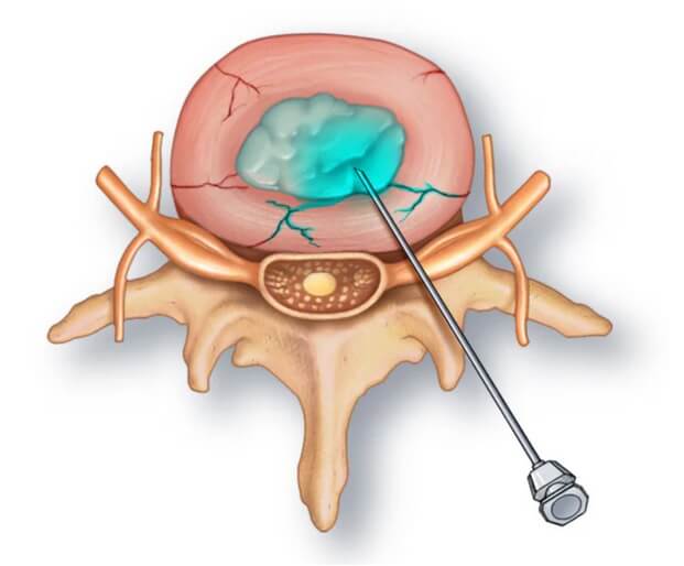
Employing fluoroscopy, an imaging technique, your doctor will monitor the entry of the discogram needle into your body. This real-time visualization ensures precise and safe placement of the needle into the center of the disc under examination. Subsequently, a contrast dye is injected into the disc, and an X-ray or CT scan is performed to observe the dispersion of the dye.
If the dye remains in the central area of the disc, it indicates normalcy. Conversely, if the dye spreads beyond the center, it suggests wear-and-tear changes in the disc. However, these changes may or may not be the direct cause of your pain.
Typically, if a disc is responsible for your back pain, you will experience pain during the injection, resembling the back pain you endure daily. On the other hand, if the disc is normal, the injection is relatively painless. Throughout the discogram, you will be prompted to articulate and rate your pain.
Typically, if a disc is responsible for your back pain, you will experience pain during the injection, resembling the back pain you endure daily. On the other hand, if the disc is normal, the injection is relatively painless. Throughout the discogram, you will be prompted to articulate and rate your pain.
Post-Procedure Care
Following the procedure, you will be under observation in the procedure room for about 30 to 60 minutes. Subsequently, you'll be permitted to leave, but it's imperative to arrange for someone to drive you home.
It's common to experience some discomfort at the injection site or in the lower back for several hours post-procedure. Applying an ice pack to the area in 20-minute intervals can help alleviate this discomfort. It's crucial to keep your back dry for the first 24 hours.

If you happen to develop severe back pain or experience a fever within one to two weeks after the procedure, it's essential to contact your doctor promptly. Immediate medical attention may be warranted in such cases.